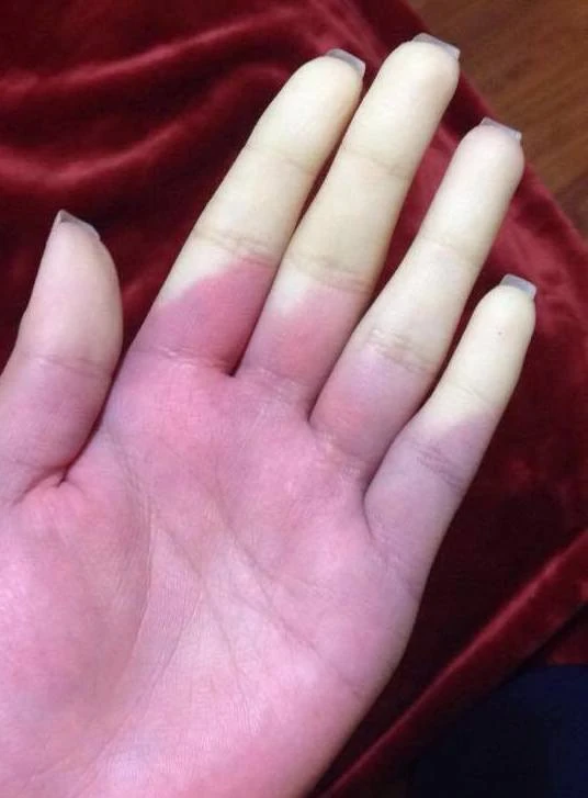
Original MICA image-reading exercise, unscored. No clinical narrative absent from the source is added.
- Describe visible findings before proposing an interpretation.
- Suggest an interpretation and at least one image limitation.
- Which clinical findings or investigations would complement this observation?
Reveal the explained reading
Raynaud phenomenon
- Trigger
- Cold or stress
- Mechanism
- Digital vasospasm
- Colours
- Pallor, cyanosis, variable redness
- Severity
- Ulcer or persistent ischaemia
Morphological description
Digits may become white, then blue and red; all three phases are not required.
Causes and diagnostic direction
Distinguish primary from secondary disease: connective-tissue disease, medicines, vibration exposure or arterial disease.
Differential diagnoses
Persistent cyanosis or fixed ischaemia warrants consideration of another circulatory disorder.
Pitfalls and limitations
An image of pallor alone establishes neither episodic behaviour nor a particular systemic disease.
Take-home message
Identify vasospasm and seek features suggesting a secondary form.
Sources
Documentary review: 2026-10-12
Media source & rights
- Author
- MSM98
- Original source
- MSM98 · Phénomène de Raynaud · https://commons.wikimedia.org/wiki/File:Raynauld.jpg · CC BY-SA 4.0
- Licence
- CC BY-SA 4.0
- Changes
- Only proportional resize and WebP conversion; no medical alterations; metadata removed.
- Dimensions
- 536 × 727 px
- Check
- 2026-10-11