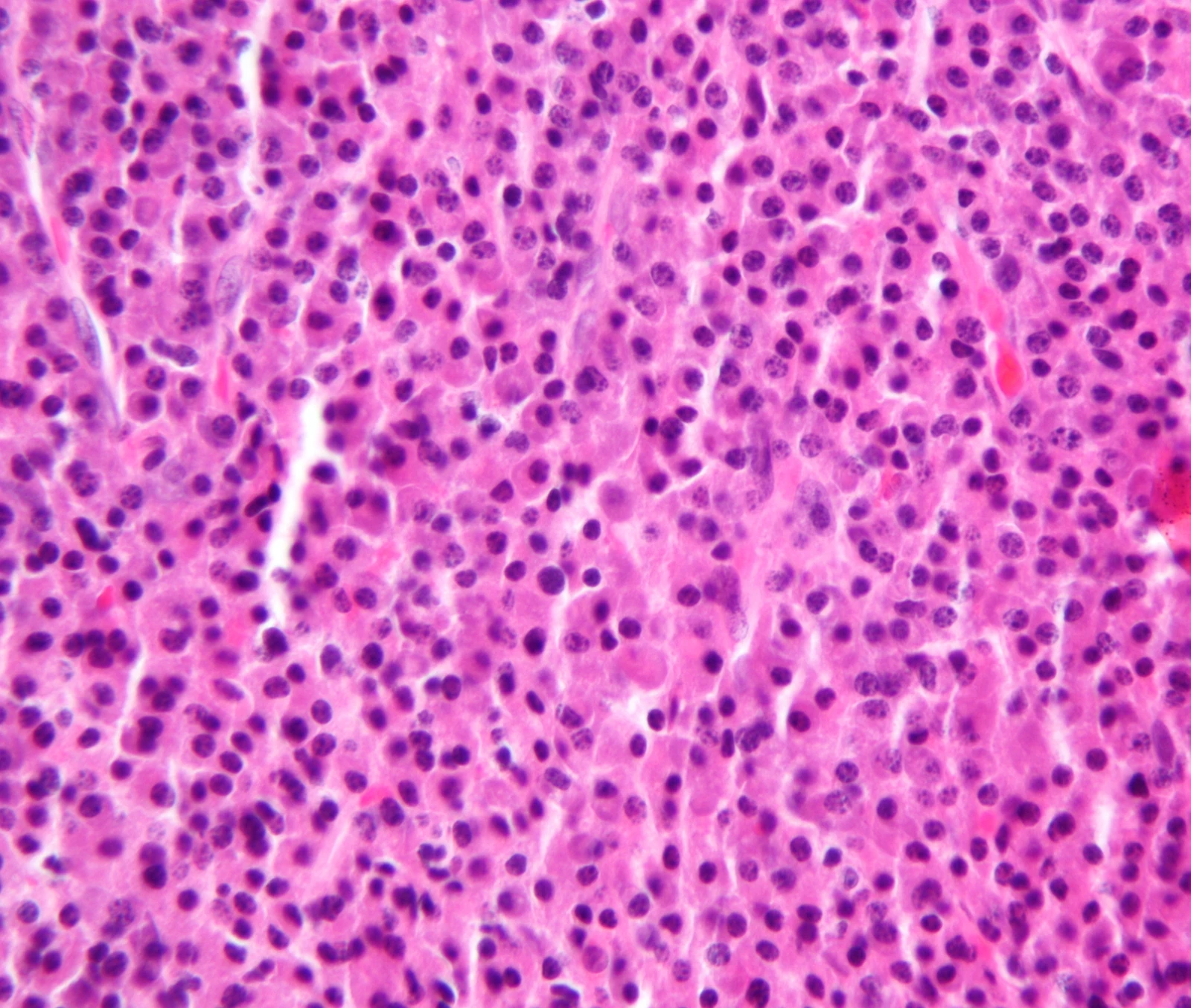
Original MICA image-reading exercise, unscored. No clinical narrative absent from the source is added.
- Describe visible findings before proposing an interpretation.
- Suggest an interpretation and at least one image limitation.
- Which clinical findings or investigations would complement this observation?
Reveal the explained reading
Plasma-cell proliferation · histology
- Cells
- Plasma cells
- Stain
- H&E
- Confirmation
- Immunophenotype and clonality
- Extent
- Marrow assessment and imaging
Morphological description
The field contains a dense plasma-cell population; nuclei are often eccentric with condensed chromatin.
Causes and diagnostic direction
A plasma-cell neoplasm may be localised or part of myeloma; the photograph does not distinguish these settings.
Differential diagnoses
Reactive plasmacytosis and other lymphoid proliferations require immunophenotypic and clonality assessment.
Pitfalls and limitations
Plasma-cell abundance alone does not prove clonality; one section cannot distinguish solitary plasmacytoma from myeloma.
Take-home message
Recognise plasma cells, confirm clonal proliferation and establish extent before classifying disease.
Sources
- Caers J et al. Diagnosis, treatment, and response assessment in solitary plasmacytoma: updated recommendations from a European Expert Panel. J Hematol Oncol, 2018 ↗
- MD Anderson Cancer Center. Solitary Plasmacytoma(Bone and Extramedullary), algorithme institutionnel, 2024 ↗
Documentary review: 2026-10-15
Media source & rights
- Author
- Nephron
- Original source
- Nephron · Plasmacytoma1.jpg · CC BY-SA 3.0 · https://commons.wikimedia.org/wiki/File:Plasmacytoma1.jpg
- Licence
- CC BY-SA 3.0
- Changes
- Full original field and annotations retained; WebP conversion, EXIF/GPS metadata removed, proportional derivatives without upscaling, cropping or medical retouching. Derivatives shared under the same CC BY-SA 3.0 licence.
- Dimensions
- 3179 × 2690 px
- Check
- 2026-10-15