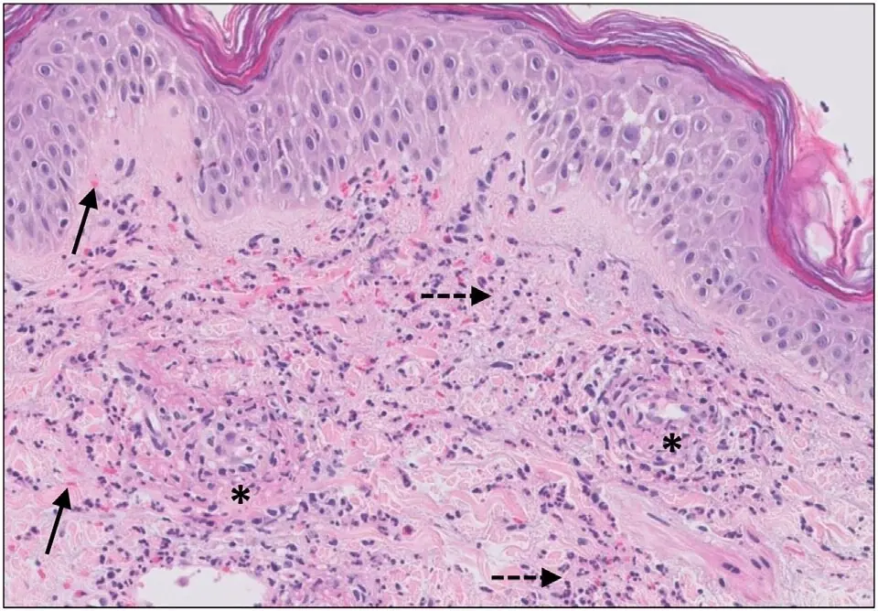
Original MICA image-reading exercise, unscored. No clinical narrative absent from the source is added.
- Describe visible findings before proposing an interpretation.
- Suggest an interpretation and at least one image limitation.
- Which clinical findings or investigations would complement this observation?
Reveal the explained reading
Leukocytoclastic vasculitis · histology
- Vessels
- Small dermal vessels
- Cells
- Neutrophils
- Debris
- Leukocytoclasia
- Wall
- Injury and fibrin
Morphological description
Assess perivascular and mural neutrophils, nuclear debris, fibrin and erythrocyte extravasation.
Causes and diagnostic direction
The pattern may follow medicines or infection, accompany systemic disease or have no identified cause.
Differential diagnoses
Capillaritis without wall destruction differs from vasculitis; isolated nuclear debris is insufficient.
Pitfalls and limitations
Histology does not always identify the cause; lesion age and specimen selection influence findings.
Take-home message
Confirm vessel injury, then distinguish skin-limited from systemic disease.
Sources
Documentary review: 2026-10-12
Media source & rights
- Author
- Jenny Giang, Marc A. J. Seelen, Martijn B. A. van Doorn, Robert Rissmann,Errol P. Prens and Jeffrey Damman
- Original source
- Jenny Giang, Marc A. J. Seelen, Martijn B. A. van Doorn, Robert Rissmann,Errol P. Prens and Jeffrey Damman — File : Micrograph of cutaneous small-vessel vasculitis.jpg — CC BY 4.0 — https://commons.wikimedia.org/wiki/File%3AMicrograph_of_cutaneous_small-vessel_vasculitis.jpg
- Licence
- CC BY 4.0
- Changes
- EXIF supprimé ; redimensionnement proportionnel ; conversion WebP ; aucun recadrage ni retouche médicale. Dérivé écran proportionnel ≤1280 px, WebP qualité76 ; vignette ≤320 px qualité72. Source acquise et dérivé initial conservés sans modification ; aucune retouche médicale, aucun recadrage.
- Dimensions
- 960 × 668 px
- Check
- 2026-10-11T20:56:34.187370+00:00