View the original document
ImagesTEACHING RESOURCE M0040
Head CT: subarachnoid haemorrhage
A CT image by Lipothymia published on Wikimedia Commons, used in the neurological station.
Lipothymia · Commons
Free MICA accountOpen
MICA
White-matter abnormalities: describe shape and location, then correlate clinically.
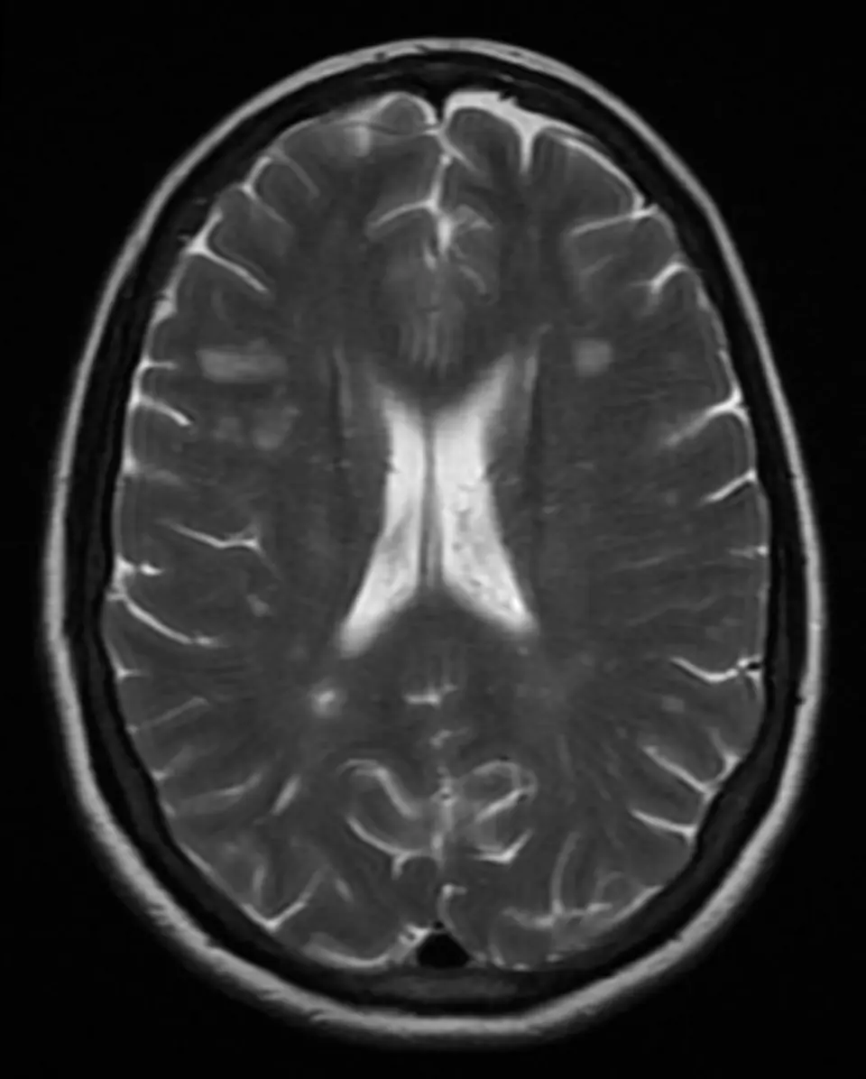
James Heilman, MD, MSMRI.png, CC BY-SA 4.0, Wikimedia Commons; conversion WebP et réduction proportionnelle.
James Heilman, MD
View the original sourceTeaching notes based on the cited sources.
TEACHING RESOURCE M0040
A CT image by Lipothymia published on Wikimedia Commons, used in the neurological station.
Lipothymia · Commons
 Images
MICA IMAGES A0070
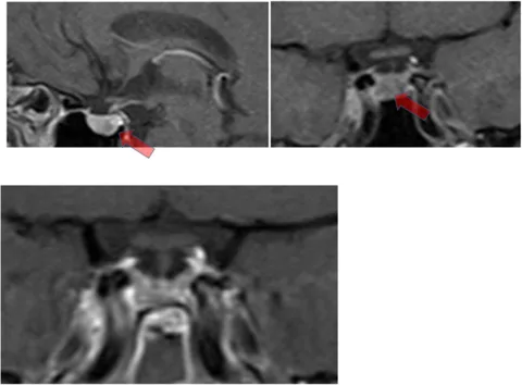
The source identifies a pituitary adenoma.
Guerreiro et al. · Figure 2 · CC BY 4.0
 Images
MICA IMAGES A0095
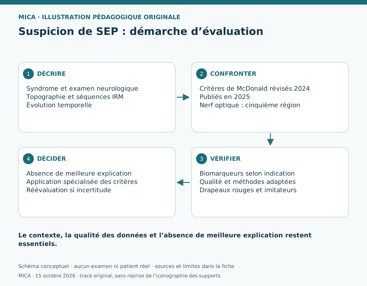
The pathway summarises questions in suspected MS. It distinguishes the revision date from the publication date.
MICA · schéma original du 15 octobre 2026 ; aucune image tierce incorporée
Learning objective
Observe and describe the document, then compare your interpretation with the atlas sheet.
Points to observe
- Identify sequence and planes, then review the whole examination and compare previous acquisitions.
Open observation mode