View the original document
ImagesTEACHING RESOURCE M0040
Head CT: subarachnoid haemorrhage
A CT image by Lipothymia published on Wikimedia Commons, used in the neurological station.
Lipothymia · Commons
Free MICA accountOpen
MICA
The source identifies a pituitary adenoma.
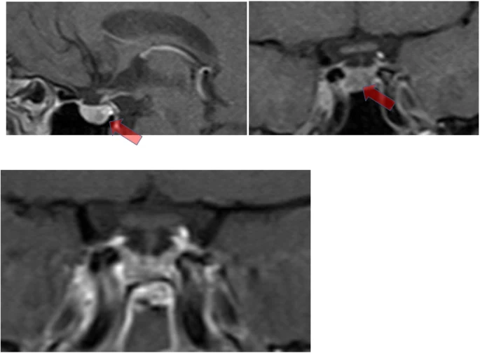
Guerreiro et al. · Figure 2 · CC BY 4.0
Guerreiro, Vanessa; Bernardes, Irene; Pereira, Josué; Silva, Roberto Pestana; Fernandes, Susana; Carvalho, Davide; Freitas, Paula
View the original sourceTeaching notes based on the cited sources.
TEACHING RESOURCE M0040
A CT image by Lipothymia published on Wikimedia Commons, used in the neurological station.
Lipothymia · Commons
 Images
MICA IMAGES A0022
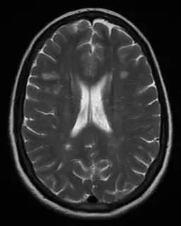
White-matter abnormalities: describe shape and location, then correlate clinically.
James Heilman, MD, MSMRI.png, CC BY-SA 4.0, Wikimedia Commons; conversion WebP et réduction proportionnelle.
 Images
MICA IMAGES A0054
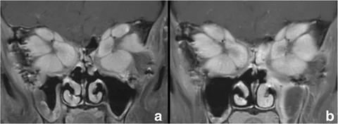
Orbital muscular involvement in a severe Graves case.
Rau, Andrea; Klopfer, Matthias; Rommel, Niklas; Rau-Fornefeld, Mechthild; Kolk, Andreas · Extraocular muscle repositioning as the last therapeutic option for a patient with a severe course of Graves’ Ophthalmopathy: a case report · CC BY 4.0 · https://link.springer.com/article/10.1186/s12886-018-0718-1
Learning objective
Observe and describe the document, then compare your interpretation with the atlas sheet.
Points to observe
- Locate the sella and arrowed lesion.
- Acromegaly diagnosis also required hormonal findings.
Open observation mode