 Images
MICA IMAGES A0042
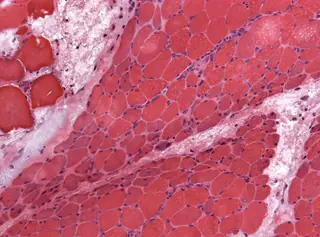
Perifascicular atrophy · muscle biopsy
Small fibres at fascicle edges: distribution is essential.
Jensflorian — File : NP MGMT 0252.jpg — CC BY-SA 4.0 — https://commons.wikimedia.org/wiki/File%3ANP_MGMT_0252.jpg
Open accessOpen
Learning objective
Observe and describe the document, then compare your interpretation with the atlas sheet.
Points to observe
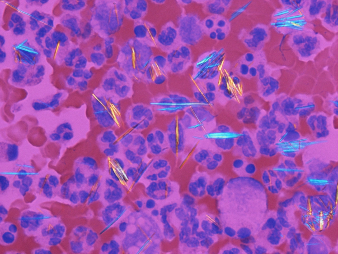
- Examine several fields and vary orientation relative to the compensator. Describe shape, intracellular or extracellular location and birefringence.
Open observation mode